Flow
Medical ultrasound is widely used to image blood flow. For instance, the estimation of blood flow velocities plays a key role in diagnosing carotid artery stenosis.
However, blood velocity estimates using conventional color flow imaging (CFI) or Doppler techniques are angle dependent. This poses a huge challenge for quantitatively measuring the magnitude (and direction) of the blood’s velocity.
The Transverse Oscillation (TO) method by Jensen and Munk remedies this fact and has shown intriguing in-vivo results of blood flow vector velocities in superficial vessels using linear arrays. It is also the technique closest to a commercial breakthrough.
However, the current implementation of the technique limits scan depth and prevents imaging of deeper lying vessels and organs, e.g. the heart and liver. Currently, the method is being expanded to a phased array for potential cardiac imaging.
To read more about our ultrasound research, please visit Center for Fast Ultrasound Imaging's website.
Imaging
Ultrasound imaging is one of the most popular diagnostic methods - it is a non-invasive and low cost technique.
However, there are certain limitations. In conventional ultrasound, the recording of images is sequential in one direction at the time. This limits how many images can be shown per second, which is especially problematic when three dimensional imaging is desired, as in scans of the heart.
At BME we are researching extensively in the ultrasound technique of the future - synthetic aperture – which should make it possible to obtain a better resolution, contrast and depth penetration.
Other research projects concentrate on methods to improve image quality by reducing speckle - the result of the constructive and destructive coherent summation of ultrasound echoes - and through optimizing operator-dependent controls.
To read more about our ultrasound research, please visit Center for Fast Ultrasound Imaging's website.
Hardware
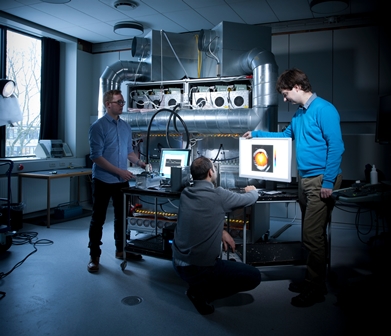
At Center for Fast Ultrasound, we are building a unique multichannel ultrasound system featuring 1024 independent channels in both transmit and receive. It will allow researchers to test new imaging techniques for better visualization of the soft tissue structures in the human body.
The data acquisition and processing logic of the system is realized using large Field Programmable Gate Array (FPGA) devices. These contain significant amounts of logic resources, which allow implementation of advanced ultrasound data processing algorithms in addition to the original ultrasound system logic.
In cooperation with its industrial partner, BK Medicial, the Center is also evaluating ultrasound imaging algorithms implemented on Graphic Processor Units (GPU), whose performance has been rapidly increasing and whose flexibility, with respect to general-purpose computing, has been steadily improving.
The algorithms being targeted for implementation are in the fields of compound imaging (speckle reduction), blood flow estimation and adaptive imaging.
To read more about our ultrasound research, please visit Center for Fast Ultrasound Imaging's website.